
Image-Guided Pain Injections: 9 Key Examples Explained

Medically reviewed by
Dr. Saurabh Dang, MD, MBA
Image-Guided Pain Injections: 9 Key Examples Explained
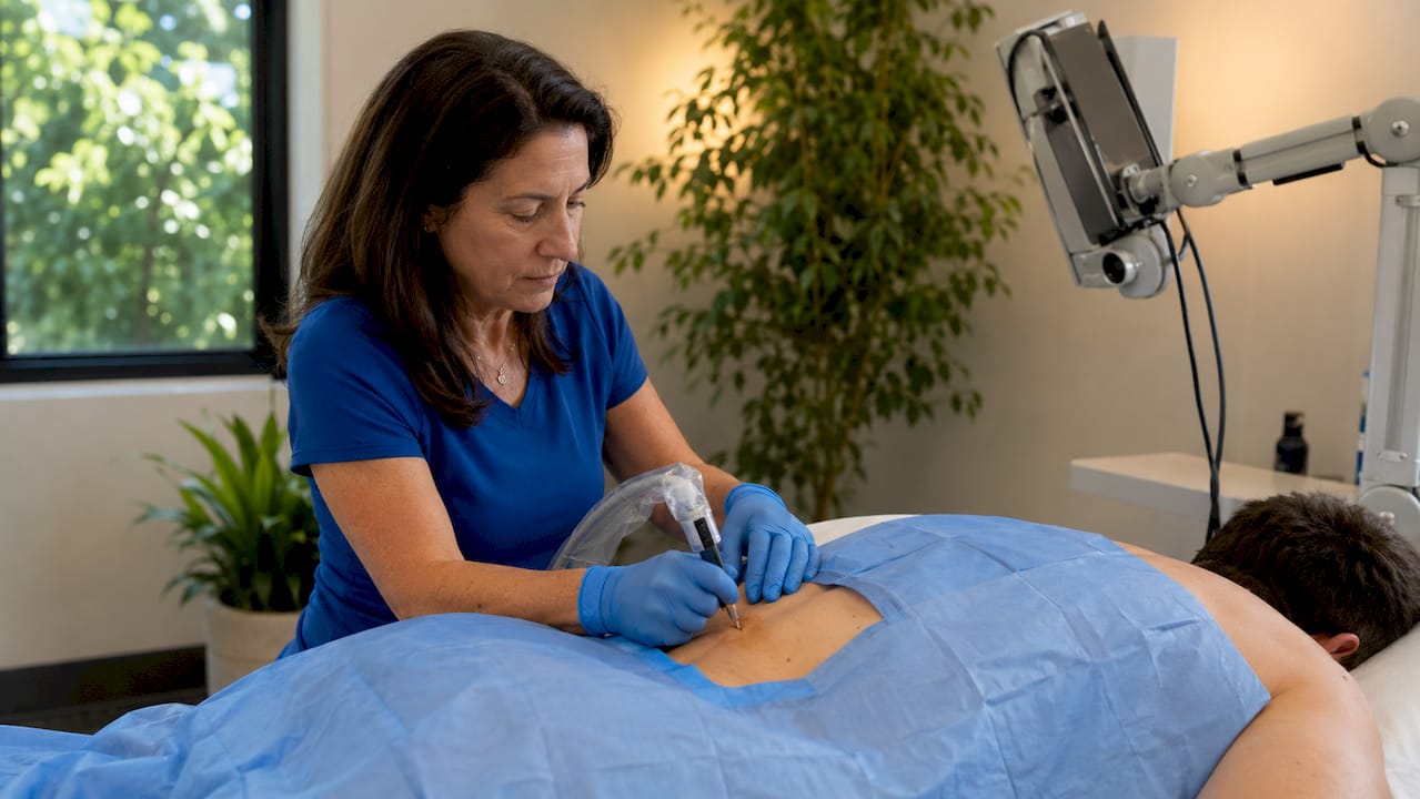
Image-guided pain injections are minimally invasive procedures that use real-time imaging, such as fluoroscopy or ultrasound, to deliver medication precisely to a targeted nerve, joint, or tissue. The industry term for this category is “image-guided interventional pain management,” and it covers a wide range of injection types. Sacroiliac joint pain alone accounts for 15–30% of chronic lower back pain cases, which shows just how many patients could benefit from targeted, guided treatment. Common examples of image-guided pain injections include epidural steroid injections, facet joint injections, nerve blocks, and ultrasound-guided joint injections. Each targets a specific pain source, and the imaging guidance is what separates these procedures from older, less accurate techniques.
1. Examples of image-guided pain injections: the most common types
The nine procedures below represent the most clinically significant examples of image-guided pain injections used in interventional pain management today. Each one uses a specific imaging method and targets a distinct anatomical structure.
Epidural steroid injections
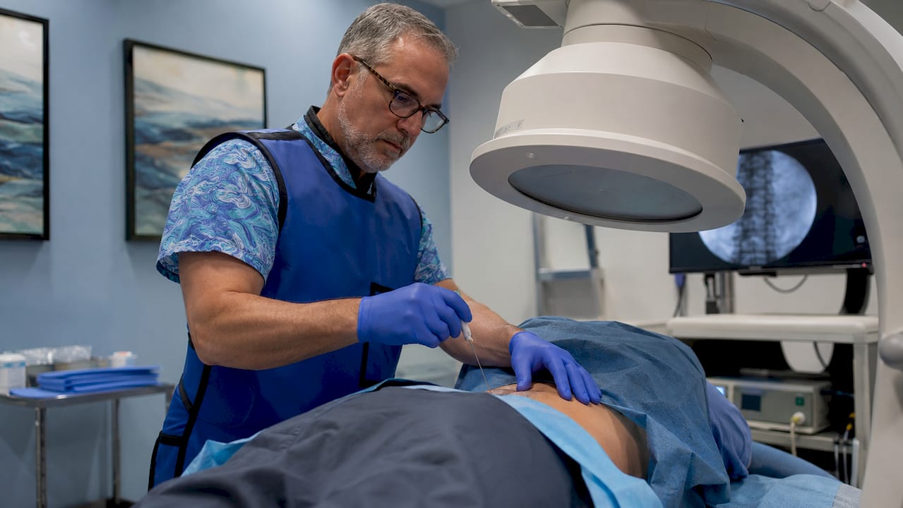
Epidural steroid injections target the epidural space surrounding the spinal cord to reduce inflammation around compressed or irritated nerve roots. Fluoroscopy guides the needle to the correct spinal level, which is critical when treating radiculopathy from a herniated disc or spinal stenosis. The injection typically contains a corticosteroid combined with a local anesthetic. Patients with sciatica or neck pain radiating into the arm are common candidates.
Facet joint injections
Facet joint injections treat arthritic or degenerative changes in the small joints connecting vertebrae in the spine. Fluoroscopy confirms needle placement inside the joint capsule before medication is delivered. These injections are especially useful for patients whose pain worsens with spinal extension or rotation. The medication typically includes a corticosteroid and a short-acting local anesthetic.
Sacroiliac joint injections
Sacroiliac (SI) joint injections address pain at the junction between the spine and the pelvis. Fluoroscopy guides the needle into the SI joint, and contrast dye confirms accurate placement before any medication is released. About 20–30% of SI joint injections reveal “leaky” joints where contrast spreads outside the capsule, requiring needle repositioning. That step protects the patient from receiving medication in the wrong tissue.
Trigger point injections
Trigger point injections treat myofascial pain syndrome, a condition where tight muscle knots cause localized and referred pain. Most trigger point injections use palpation alone, but ultrasound guidance adds value when the target muscle is deep or the patient’s anatomy makes surface landmarks unreliable. A small volume of local anesthetic, sometimes combined with a corticosteroid, is injected directly into the knot. Relief can be immediate and lasting when the correct spot is targeted.
Ultrasound-guided knee injections
Knee injections under ultrasound guidance deliver corticosteroids or platelet-rich plasma (PRP) directly into the joint space for osteoarthritis or inflammatory conditions. Ultrasound allows direct visualization of fluid pockets before needle insertion, which improves accuracy and reduces patient discomfort. This is particularly helpful in patients with high body mass index, where surface landmarks are difficult to locate. The real-time view also confirms the needle tip is inside the joint before injection.
Pro Tip: When a knee joint contains excess fluid, use the IV catheter technique under ultrasound to drain fluid and inject medication through a single access point. This reduces the number of needle sticks and significantly improves patient comfort.
Shoulder injections
Ultrasound-guided shoulder injections target the glenohumeral joint, subacromial bursa, or biceps tendon sheath depending on the diagnosis. Rotator cuff tears, adhesive capsulitis, and bursitis all respond to precisely placed corticosteroid injections. Ultrasound eliminates the guesswork of blind injection techniques, which carry a meaningful risk of missing the target structure entirely. PRP is also used in this location for patients seeking a regenerative approach.
Hip joint injections
Hip injections require careful targeting because the joint sits deep beneath muscle and fat. Anatomical variation means the needle must target the lateral femoral head-neck junction to avoid accidentally entering the bursa instead of the joint. Fluoroscopy or ultrasound confirms placement before the physician injects the therapeutic mixture. Patients with hip osteoarthritis or labral pathology are the most common candidates.
Nerve blocks
Image-guided nerve blocks interrupt pain signals along a specific nerve pathway using a local anesthetic, a corticosteroid, or both. They serve two distinct roles: diagnostic blocks confirm whether a specific nerve is the pain source, and therapeutic blocks provide lasting relief. Nerve blocks offer a non-opioid alternative for patients with chronic pain who want to avoid or reduce medication dependence. Fluoroscopy or ultrasound guides the needle to the correct nerve location with precision.
Platelet-rich plasma injections
PRP injections use a concentration of the patient’s own platelets to promote tissue healing in damaged joints, tendons, and ligaments. Ultrasound guidance confirms the needle reaches the exact site of injury, which is critical because PRP works by stimulating local cellular repair. This approach suits patients with early-stage osteoarthritis or chronic tendon injuries who have not responded to corticosteroid injections. PRP represents the regenerative end of the image-guided injection spectrum.
2. How imaging guidance improves safety and effectiveness
Real-time imaging changes the outcome of pain injections in ways that matter to patients. The core benefit is direct visualization: the physician sees the needle tip moving toward the target structure and can correct course before any medication is delivered.
Accuracy and first-attempt success
Ultrasound guidance improves first-attempt success rates compared to landmark-based methods, which reduces the number of needle passes and the associated discomfort. Fewer attempts also mean less tissue trauma and a lower risk of post-procedure soreness. For patients who are anxious about injections, a faster and more accurate procedure makes a real difference.
Radiation and patient safety
Ultrasound produces no radiation, making it the preferred modality for office-based procedures and for patients who need repeated injections over time. Fluoroscopy uses low-dose X-ray and remains the standard for spinal procedures where bony landmarks must be confirmed. CT guidance offers the highest anatomical detail but is reserved for complex cases because of higher radiation exposure and cost.
| Imaging modality | Radiation exposure | Best suited for |
|---|---|---|
| Ultrasound | None | Joint, soft tissue, and office-based injections |
| Fluoroscopy | Low dose X-ray | Spinal injections, SI joint, facet joint |
| CT guidance | Higher dose X-ray | Complex anatomy, failed prior injections |
Pro Tip: Ask your physician whether ultrasound guidance is an option for your injection. For many joint and soft tissue procedures, ultrasound delivers the same accuracy as fluoroscopy with zero radiation exposure.
Contrast confirmation
Contrast dye under fluoroscopy confirms needle placement by showing how the injected fluid spreads within the target structure. If the contrast pattern looks wrong, the physician repositions the needle before delivering the therapeutic medication. This step is standard for spinal and SI joint injections and is one of the clearest examples of how imaging protects the patient.
3. Key medications used in image-guided injections
The medication delivered through an image-guided injection determines the type and duration of pain relief. Physicians select the agent based on the diagnosis, the target structure, and the patient’s treatment history.
- Local anesthetics such as lidocaine 2% provide immediate pain relief and serve a diagnostic function. If the pain disappears after injection, that confirms the targeted structure is the pain source.
- Corticosteroids reduce inflammation at the injection site and provide longer-lasting relief, typically measured in weeks to months. They are the most commonly used therapeutic agent in spinal and joint injections.
- Platelet-rich plasma (PRP) promotes tissue healing by concentrating growth factors from the patient’s own blood. It is used when the goal is repair rather than symptom suppression.
- Combination mixtures of anesthetic and corticosteroid are standard for most joint and spinal injections, with total volumes typically ranging from 1.0–2.0 mL to avoid tissue distention and post-injection pain.
Rapid medication infusion can cause pain from tissue distention, so physicians inject slowly and monitor patient feedback throughout the procedure. Topical anesthetics applied to the skin before needle insertion also reduce discomfort at the entry point. Controlling volume and injection speed is as important as choosing the right medication.
4. Which injection type fits which condition?
Choosing the right injection depends on where the pain originates and what the imaging shows. Spinal injections are site-specific, meaning the same symptom, such as lower back pain, can require a different injection depending on whether the source is a disc, a facet joint, or the SI joint.
| Condition | Recommended injection type | Imaging modality |
|---|---|---|
| Herniated disc with radiculopathy | Epidural steroid injection | Fluoroscopy |
| Facet joint arthritis | Facet joint injection or medial branch block | Fluoroscopy |
| Sacroiliac joint dysfunction | SI joint injection | Fluoroscopy with contrast |
| Knee or shoulder osteoarthritis | Intra-articular corticosteroid or PRP | Ultrasound |
| Myofascial pain or muscle knots | Trigger point injection | Palpation or ultrasound |
| Chronic nerve pain | Diagnostic or therapeutic nerve block | Fluoroscopy or ultrasound |
Patients with high body mass index benefit most from ultrasound guidance because surface landmarks become unreliable when soft tissue depth increases. Office-based ultrasound also removes the need for a radiology suite, which makes scheduling faster and more convenient. For complex spinal anatomy or prior surgical history, fluoroscopy with contrast remains the most reliable choice.
Key takeaways
Image-guided pain injections are the most effective minimally invasive option for chronic joint, spinal, and nerve pain because real-time imaging confirms needle placement before any medication is delivered.
| Point | Details |
|---|---|
| Imaging modality matters | Choose ultrasound for zero radiation in joints; use fluoroscopy for spinal and SI joint procedures. |
| SI joint pain is common | Sacroiliac dysfunction accounts for 15–30% of chronic lower back pain cases. |
| Contrast confirms accuracy | Fluoroscopy with contrast dye catches “leaky” joints in 20–30% of SI injections. |
| Medication volume is critical | Keep total injection volume at 1.0–2.0 mL to avoid tissue distention and post-procedure pain. |
| Nerve blocks serve two roles | A single nerve block can both diagnose the pain source and deliver therapeutic relief. |
What I’ve learned watching image-guided injections evolve
The shift toward ultrasound in clinic settings is the most significant change I have seen in interventional pain management over the past decade. Wireless ultrasound scanners have removed the last practical barrier to office-based guided injections. Physicians no longer need a fluoroscopy suite to deliver accurate, image-confirmed treatment.
What surprises most patients is how much the imaging changes the conversation before the needle even enters the skin. Seeing a fluid pocket in a knee joint or a thickened bursa in a shoulder on the ultrasound screen gives both the physician and the patient a shared understanding of what is happening. That visual confirmation builds trust in a way that a verbal explanation alone cannot.
The non-opioid angle is also worth stating plainly. Image-guided nerve blocks support opioid-reduction strategies in chronic pain management by providing targeted relief without systemic medication. For patients who are trying to reduce or avoid opioids, a well-placed nerve block or epidural injection can be the difference between managing and not managing their pain.
My one caution: not every injection is appropriate for every patient, and anatomical variation is real. A physician who skips the contrast step in an SI joint injection or who does not account for depth in a hip injection is taking a shortcut that costs the patient. Ask your provider whether they use imaging guidance and contrast confirmation. Those two questions tell you a great deal about the quality of care you are about to receive.
Pain management at Hudson Pain and Spine in Northern New Jersey
Hudson Pain and Spine serves patients across Bergen, Passaic, and Middlesex counties with a full range of image-guided injection therapies, including epidural steroid injections, nerve blocks, facet joint injections, and SI joint procedures.
If you are living with chronic back pain, sciatica, joint pain, or nerve-related discomfort, a consultation can help identify which injection type fits your specific condition. Visit pain management services to review available treatments and book an appointment at a location convenient to you across Northern and Central New Jersey.
FAQ
What are image-guided pain injections?
Image-guided pain injections are minimally invasive procedures that use fluoroscopy or ultrasound to deliver medication precisely to a targeted nerve, joint, or spinal structure. Real-time imaging confirms needle placement before any medication is released.
How long does pain relief last after a guided injection?
Relief duration varies by injection type and individual response. Corticosteroid injections typically provide relief lasting several weeks to months, while PRP injections aim for longer-term tissue repair.
Are image-guided injections safer than blind injections?
Yes. Direct visualization reduces the risk of injecting outside the target structure and improves first-attempt accuracy, which lowers patient discomfort and the chance of complications.
Which imaging method is best for joint injections?
Ultrasound is the preferred modality for peripheral joint injections such as the knee, shoulder, and hip because it produces no radiation, allows real-time visualization of fluid, and works well in an office setting.
Can image-guided injections replace opioid medications?
Image-guided nerve blocks and spinal injections are a recognized non-opioid pain management strategy. They do not replace all medications for every patient, but they can significantly reduce or eliminate the need for opioids in many chronic pain cases.
Recommended
- Best Injections for Chronic Knee Pain | Hudson Pain and Spine Blog
- Epidural Steroid Injections: What to Expect Before, During, and After | Hudson Pain and Spine Blog
- Pain Management Blog | Hudson Pain and Spine NJ
- Epidural Injections in Englewood, NJ | Hudson Pain and Spine
About the Medical Reviewer
Dr. Saurabh Dang is a double board-certified interventional pain management specialist serving Central and Northern New Jersey. He combines clinical expertise with a patient-centered approach to help patients find lasting relief from chronic pain conditions.
Read Full Bio →Seeking Treatment for Epidural Injections?
Dr. Dang and the team at Hudson Pain and Spine offer specialized care and advanced interventional treatments.
Ready to Find Relief from Pain?
Schedule your consultation with Dr. Saurabh Dang at our Englewood office.
Serving patients across Central and Northern New Jersey — Bergen, Passaic, and Middlesex counties.