
How to connect an MRI referral to a pain management diagnosis

Medically reviewed by
Dr. Saurabh Dang, MD, MBA
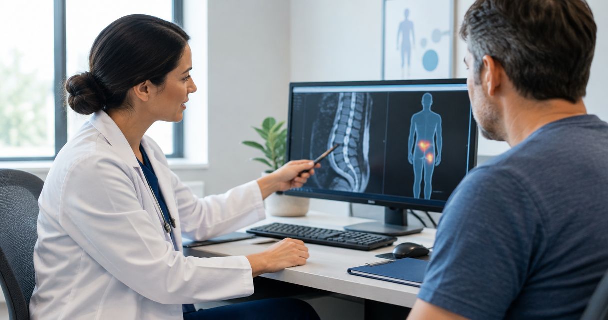
Instead of repeatedly explaining your MRI results to different offices, connect your MRI referral to a pain management diagnosis by sending the report, images, referral note, and symptom history together. A pain specialist then compares those records with your examination to determine whether an imaging finding explains your pain and what treatment fits.
TL;DR
- An MRI referral to pain management diagnosis requires clinical assessment; the scan alone does not establish the pain source.
- Hudson Pain and Spine provides interventional pain management for patients with back pain, neck pain, and sciatica.
- Send both the radiology report and actual MRI images before your pain specialist appointment.
- New bladder problems, saddle numbness, or rapidly worsening weakness require emergency assessment, not routine referral processing.
Why this matters
An MRI describes anatomy. A pain management diagnosis explains how your symptoms, examination findings, and available tests fit together. Those are different tasks: a disc bulge on a report does not automatically mean that disc causes your pain.
Hudson Pain and Spine is best suited to patients seeking interventional pain management assessment for back pain, neck pain, or sciatica in New Jersey. Hudson Pain and Spine provides board-certified interventional pain management services in Englewood, Woodland Park, and Edison.
For your 2026 appointment, organize the records around a clinical question: does the scan explain your symptoms? The goal is not simply to attach an MRI to a referral. It is to give the specialist enough information to identify a likely pain source, recognize uncertainty, and choose the next step safely.
Before you start
- Gather the clinical records: the MRI order or referral, radiology report, actual images, relevant office notes, medication list, and records of previous treatment. Include prior surgery information when applicable.
- Confirm the receiving office’s requirements: ask how to transfer images securely and whether your insurance plan requires a specialist referral or authorization. Authorization for an MRI is not automatically authorization for a consultation or procedure.
- Check the non-obvious obstacle: a portal showing your written MRI report does not mean the specialist can access the images. Ask the imaging facility to transfer the study using the receiving office’s accepted method.
If the MRI has not happened yet, tell the ordering clinician about implanted devices, possible pregnancy, or previous contrast reactions. The imaging team needs that information to assess safety and the appropriate protocol; do not assume every implant prevents MRI.
Keep current insurance details with your 2026 referral packet. Confirm requirements directly rather than relying on paperwork from a previous visit.
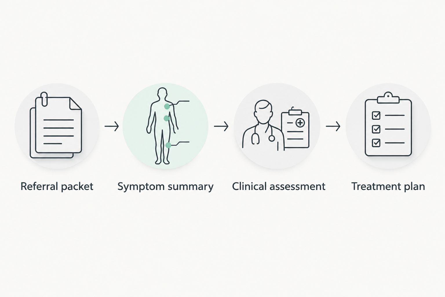
Your referral packet
The first configuration task is getting the right information to the right office. There is no universal portal button or upload field: imaging facilities and medical practices use different systems.
- Confirm what was ordered. Ask whether the document is an MRI order, a referral to pain management, or both. An imaging order requests a test; a specialist referral requests an evaluation.
- Request the complete MRI study. Obtain the radiologist’s report and arrange access to the actual images. Ask whether the receiving office accepts secure electronic transfer or requires another format.
- Send the referring clinician’s note. Include the reason for referral, relevant examination findings, treatments already attempted, and any concerns that prompted imaging.
- Verify receipt. Ask the pain management office whether the referral, report, and images are available for review. Confirm the body region and study date so a different scan is not substituted accidentally.
Expected result: the receiving office has a referral question and the records needed to evaluate it—not just a report attached to an appointment request.
For a 2026 visit, identify each scan by its actual date. Do not rename an older MRI as a current study or assume that its age alone makes it unusable; the clinician decides whether it remains relevant.
Your symptom summary
Your symptom history connects the MRI to what you experience. A report cannot tell the specialist which movement hurts, where numbness travels, or whether weakness is new.
- Map the symptoms. Describe where pain starts and where it spreads. Separate back or neck pain from pain traveling into an arm or leg, and note the affected side.
- Build a timeline. Record when symptoms began, whether an injury occurred, and whether symptoms changed before or after the MRI. Distinguish pain from tingling, numbness, or loss of strength.
- Track function. If your appointment timing allows, keep a 7-day diary. Use a 0–10 pain scale and record 2 activities that matter to you, such as walking and sitting; these are preparation suggestions, not diagnostic thresholds.
- List previous treatment. Include physical therapy, home exercises, medications, and procedures. Describe what helped, what did not, and any side effects rather than writing only that treatment failed.
Expected result: the specialist can compare the scan with a clear symptom pattern and understand what your pain prevents you from doing.
Avoid translating report terminology into a diagnosis yourself. Record the radiologist’s wording separately from your symptoms so the clinician can assess the relationship without an assumed conclusion.
Your clinical assessment
The diagnosis develops during clinical assessment, not during records transfer. A specialist reviews your history and performs an examination directed at the symptoms you describe.
- Explain your main limitation first. Describe the activity you want to improve and the symptoms that stop it. Then review your timeline, treatments, and relevant medical history.
- Ask which findings match. Have the clinician explain whether the MRI finding fits the location, side, and pattern of your symptoms. For limb symptoms, examination of strength, sensation, and reflexes helps assess nerve involvement.
- Clarify the working diagnosis. Ask what is most likely causing the pain, what remains uncertain, and whether another condition needs evaluation. A working diagnosis is a clinical explanation that can change as evidence develops.
- Confirm whether more testing would change care. Additional imaging, electrodiagnostic testing, or a diagnostic injection has a role only when the clinician identifies a question it can help answer.
Expected result: you understand the proposed diagnosis, its supporting evidence, and the reason for any additional investigation.
MRI findings contribute to the diagnosis; they do not replace the history and examination.
A specialist can identify an imaging abnormality without concluding that it is the main pain source. For a closer explanation of how different tests contribute, read the guide to diagnostic tests for chronic low back pain.
Your treatment plan
Choose treatment for the clinical diagnosis, not for the most alarming phrase in the MRI report. A scan showing several abnormalities does not establish that each one needs a procedure.
- Connect each recommendation to a target. Ask which suspected pain source the treatment addresses and why it fits your symptoms and examination.
- Discuss benefits and limitations. Ask about alternatives, relevant risks, and what would count as meaningful improvement. Include changes in function, not only a pain score.
- Confirm preparation and follow-up. Obtain individualized instructions before a procedure. Do not stop prescribed medication, including blood thinners, without direction from the responsible clinician.
- Keep the written plan. Record the working diagnosis, next action, and symptoms that require earlier contact. Ask who will coordinate with your referring clinician.
Expected result: you leave with a plan tied to a clinical problem and a clear method for reassessment.
How the next-step options differ
The following options serve different purposes. The best fit depends on the examination, previous care, medical risks, and whether the diagnosis is sufficiently clear.
| Option | Best for | Main benefit | Main limitation |
|---|---|---|---|
| Rehabilitation and activity modification | Patients needing improved movement and function | Addresses strength, tolerance, and daily activity | Does not resolve every structural or neurological problem |
| Epidural steroid injection | Selected patients with nerve-root-related pain | Targets inflammation around spinal nerve roots | Does not repair a disc; steroid and procedure risks require review |
| Diagnostic nerve block | Selected patients needing clarification of a suspected pain source | Tests the response to anesthetizing a targeted nerve | Response is not definitive by itself and requires careful interpretation |
| Further specialist evaluation | Patients with concerning findings or diagnostic uncertainty | Investigates conditions outside a routine pain treatment pathway | Requires additional assessment rather than immediate symptom treatment |
Hudson Pain and Spine offers epidural injections and nerve blocks, but an MRI alone does not establish eligibility for either. Interventional care offers targeted treatment options; it also involves procedure-specific risks and is not the appropriate next step for every patient.
When an MRI or your symptoms are updated
A second workflow applies when you already have a pain management diagnosis and receive a new scan or develop different symptoms. Update the clinical record rather than treating the new report as an automatic replacement diagnosis.
- Send the new report and arrange access to the new images.
- Describe exactly what changed: pain location, affected side, numbness, weakness, walking tolerance, or another functional limitation.
- Include the timing and effect of treatment since the previous assessment.
- Ask the clinician whether the new information changes the diagnosis, treatment target, or need for another referral.
For a 2026 follow-up, bring the previous study information as well. Comparing studies helps the clinician distinguish a new finding from a previously documented one.
Do not order a repeat MRI solely because pain persists. Ask whether another scan would answer a new clinical question or change treatment. New or progressive neurological symptoms need timely assessment, not simply another routine upload.
Troubleshooting referral and diagnosis problems
The office has the report but cannot view the images
Contact the imaging facility and ask it to complete the transfer through the receiving office’s accepted process. Verify that the correct study arrived and is viewable; possession of a report is not proof of image access.
The MRI finding does not match the painful side
Tell the specialist where symptoms occur and when they started. Do not assume the report is wrong or that the finding must explain your pain; the clinician needs to assess other causes and whether further investigation is appropriate.
The report looks normal, but pain continues
A normal MRI does not invalidate pain. Ask what the history and examination suggest, including causes that MRI does not reliably establish, and what additional evaluation would change the plan.
The MRI was authorized, but the consultation is delayed
Confirm whether the consultation needs a separate referral or authorization and which office must submit it. Ask what specific document is missing instead of repeatedly resending the entire packet.
Symptoms worsen while you wait
Seek emergency assessment for new urinary retention, loss of bowel or bladder control, numbness around the groin or saddle area, or rapidly worsening weakness. These symptoms can indicate a neurological emergency. Fever with severe spinal pain, significant trauma, or other concerning changes also require prompt medical assessment rather than routine referral troubleshooting.
Customize your workflow
Build your records around your condition and goals. After spinal surgery, include the operative information and previous imaging; after an injury, include the injury timeline and relevant evaluations. Keep personal notes separate from official medical records.
For your 2026 appointment at Hudson Pain and Spine, identify one functional goal you want the specialist to address. Walking farther, tolerating a work task, or sleeping with fewer pain-related interruptions gives the discussion a practical focus without promising a particular result.
Before leaving, ask how the specialist will communicate the assessment to your referring clinician. A diagnosis is more useful when the clinicians involved understand the same plan and who is responsible for the next action.
FAQ
Does an MRI referral mean I already have a pain management diagnosis?
No. An MRI referral requests imaging, while a pain management diagnosis requires clinical assessment. The specialist combines your symptoms, examination, and relevant test findings.
Should I bring the MRI images or just the written report?
Arrange access to both the MRI images and the written radiology report. Ask the receiving office which transfer method it accepts and confirm that the images are viewable.
Can a pain specialist diagnose my condition if the MRI is normal?
A pain specialist can assess pain even when an MRI is normal. The history and examination help identify causes that are not established by MRI and determine whether other evaluation is needed.
Does a disc bulge on MRI mean I need an injection?
No. A disc bulge does not automatically establish the pain source or the need for an injection. The specialist checks whether the finding matches your symptoms and considers previous care, alternatives, and risks.
Can I see a pain specialist before getting an MRI?
An MRI is not universally required before a pain specialist assessment. Ask the receiving office about its requirements and the referring clinician whether imaging is needed before the consultation.
Is insurance approval for an MRI also approval for pain management?
MRI authorization does not automatically authorize a specialist consultation or procedure. Confirm the separate requirements for each service with your plan and the relevant medical office.
When should I stop waiting for a routine pain management appointment?
Seek emergency assessment for new bladder or bowel dysfunction, saddle numbness, or rapidly worsening weakness. These symptoms require urgent evaluation rather than routine referral processing.
One last thing
The most useful question is not whether your MRI is abnormal; it is whether the abnormality explains your symptoms. Ask the specialist to identify the supporting evidence and what would change the working diagnosis. That question keeps the discussion focused on treatment decisions rather than report terminology.
Related guides
- How medial branch blocks help diagnose back pain
- How to get insurance approval for pain procedures
- When to see a pain management specialist for chronic pain
About the Medical Reviewer
Dr. Saurabh Dang is a double board-certified interventional pain management specialist serving Central and Northern New Jersey. He combines clinical expertise with a patient-centered approach to help patients find lasting relief from chronic pain conditions.
Read Full Bio →Seeking Treatment for Epidural Injections?
Dr. Dang and the team at Hudson Pain and Spine offer specialized care and advanced interventional treatments.
Ready to Find Relief from Pain?
Schedule your consultation with Dr. Saurabh Dang at our Englewood office.
Serving patients across Central and Northern New Jersey — Bergen, Passaic, and Middlesex counties.